Molecular Microscopy: Italian-Israeli team develops a new technique to study biological tissues

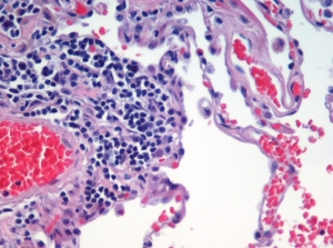
Emphysema H and E
Biological tissues are composed of millions of cells of different kinds, like enormous puzzles. While scientists can identify which kind of cells are there, they have not been able to determine how they are arranged within tissues. A study published a few days ago in Science might change everything.
Researchers in the Dynamics of Immune Responses Unit at Ospedale San Raffaele, Italy, guided by Matteo Iannacone, have joined forces with the teams of Ido Amit and Ziv Shulman at the Weizmann Institute of Science, Israel, to create a powerful and innovative technique to help address this problem.
The scientists have combined intravital microscopy (which allows them to follow the movements of cells in the tissues) and the analysis of gene expression, and have managed to map how different cells are arranged in tissues. The study has its roots in the interest of the three scientists in the dynamics of immune responses. Our immune cells move around our body and react according to biochemical signals sent by the tissues they reach. That is why understanding our immune system behavior is essential to analyze the tissue and the cells they comprise.
This study was funded by the Armenise-Harvard Foundation, the European Research Council (ERC), the Italian Association for Cancer Research (AIRC), the Italian Ministry of Health and the Lombardy Foundation for Biomedical Research.
Find out more


